Molecular connectomics

Industrializingthe connectome.

panluminate develops the chemistry, instruments, and software that make synaptic-resolution brain mapping possible at scale. Our Signature Service puts that capability to work on your tissue today.

Explore the platform
Connectome reconstruction from panluminate pan-ExM data

Trusted by leading research institutions

Harvard UniversityYale UniversityAllen InstituteUniversity of Southern CaliforniaNational Institute of Allergy and Infectious Diseases
The platform

Expand. Image. Map.

One workflow, from tissue to connectome. Each step has its own instrument, designed to work with the others. panluminate is the only company developing all three.

panexpanse™ Waitlist

Tissue expansion, automated.

panexpanse runs pan-ExM sample preparation without an operator at the bench, so every sample expands and pan-stains the same way, batch after batch.

About 20×linear tissue expansion
Hands-offautomated pan-ExM protocol
Reproduciblefrom batch to batch

Running pan-ExM at your own bench? Our pan-ExM kits bring the same chemistry to your lab.
panoptix™ Early access

A light-sheet microscope built for expanded tissue.

panoptix images large expanded samples in near-isotropic 3D, with a sample chamber designed around pan-ExM. Jointly developed with 3i.

Nanoscaleeffective resolution after expansion
Near-isotropicvolumetric imaging
pan-ExM readypurpose-built sample chamber

cortex™ Waitlist

From image to connectome.

cortex is our AI-native platform for molecular connectomics. It brings imaging data, dense segmentation, synapse detection, and molecular labels together so you can explore circuits and the molecules within them.

Dense segmentationevery neuron in the volume
0.94 F1AI-assisted presynapse detection
8 channelsmolecular labels co-registered

Dense segmentation model co-developed with Scalable Minds. Powered by WEBKNOSSOS.

Dense reconstruction of panluminate pan-ExM data
The science underneath

Wiring and molecular identity, together

pan-Expansion Microscopy (pan-ExM) physically enlarges tissue about 20-fold and stains all of its protein. A light microscope can then resolve individual synapses and up to eight molecular targets in the same sample.

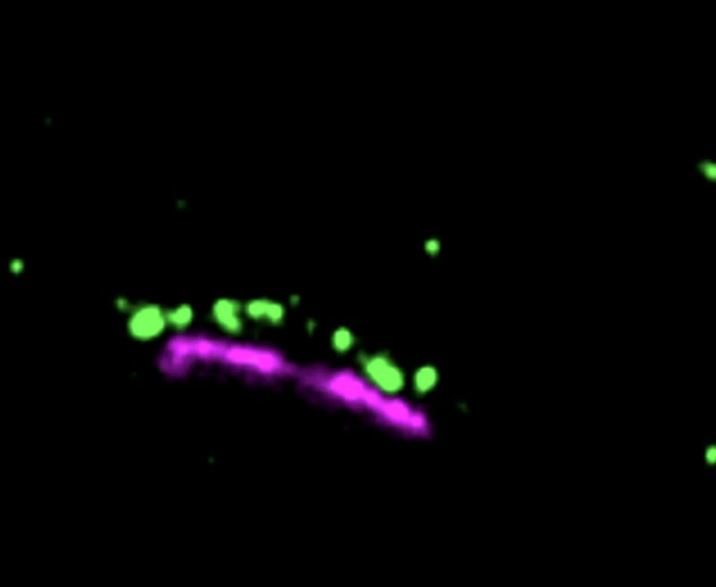
  1. Synaptic markers shown as green and magenta points on black

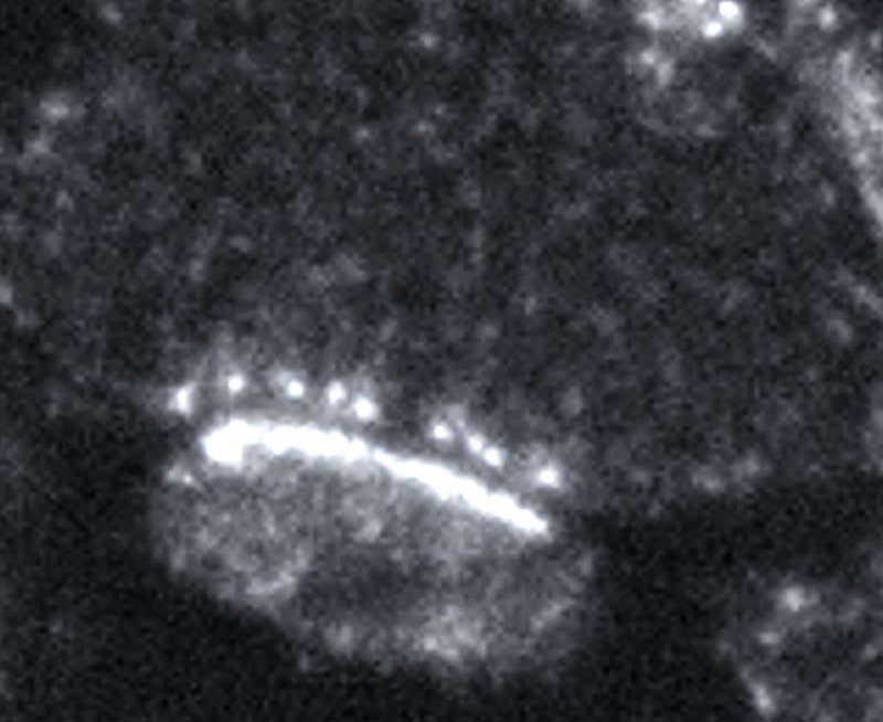
    Fluorescence alone

    Labeled proteins appear as isolated points, with no structure around them.

  2. The same synapse in grayscale pan-stain

    pan-staining

    Every synapse, membrane, and organelle becomes visible, with detail approaching electron microscopy.

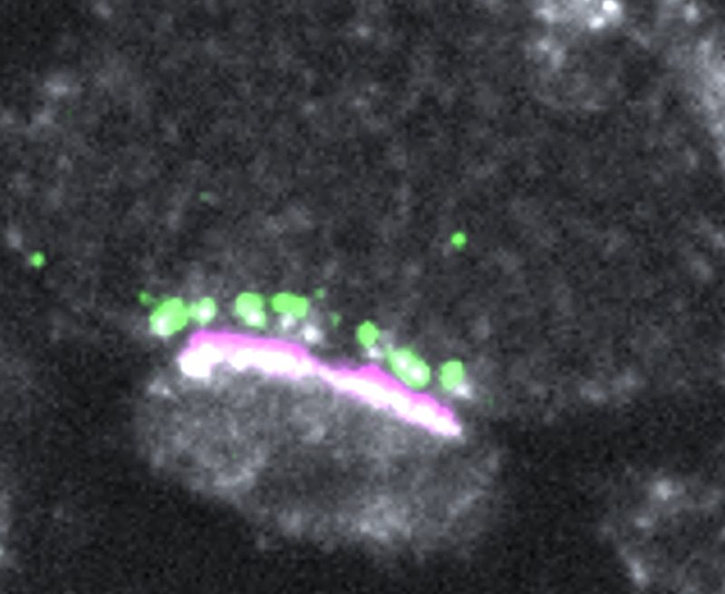
  3. Synaptic markers overlaid on the pan-stained synapse

    Molecules in context

    Specific proteins located precisely within their structural surroundings.

  4. AI-assisted analysis

    AI reconstructs neurons in 3D from pan-stained volumes, using molecular signals to pinpoint their synapses.

    Segmentation powered by WEBKNOSSOS, co-developed with Scalable Minds.

panluminate Signature Service

Or let our team run it for you.

Send us your tissue. The scientists who invented pan-ExM take it from study design through imaging data delivery, for academic labs, core facilities, pharmaceutical teams, and NeuroAI companies.

  1. 01Study design consultation with our scientists
  2. 02Sample preparation and expansion with our patented pan-ExM chemistry
  3. 03Multiplexed labeling of up to eight targets
  4. 04High-resolution volumetric imaging
  5. 05Imaging data delivered ready for your analysis pipeline
Species5 and counting
  • Human
  • Mouse
  • Fish
  • Frog
  • Fruit fly
  • More
Tissues7 and counting
  • Brain
  • Retina
  • Heart
  • Lung
  • Liver
  • Kidney
  • Intestine
  • More

Our team has already expanded and imaged every one of these. More includes olfactory tissue, testes, and human postmortem tissue.

Available now
Who we work with

Built for every team mapping the brain

Academic labs and core facilities

Synaptic-resolution imaging with molecular labeling, without building the method in-house.

Founding Core Program

Pharma and CNS drug discovery

See how disease models and treatments change synapses, and the molecules within them, in the same tissue.

NeuroAI and brain-inspired computing

Ground-truth data on how real neurons connect, and what they're made of.

NeuroAI Partner Program

Ready to mapat scale?

Tell us about your tissue and your research question. We'll respond within two business days.