
Image
panoptix™
Available now
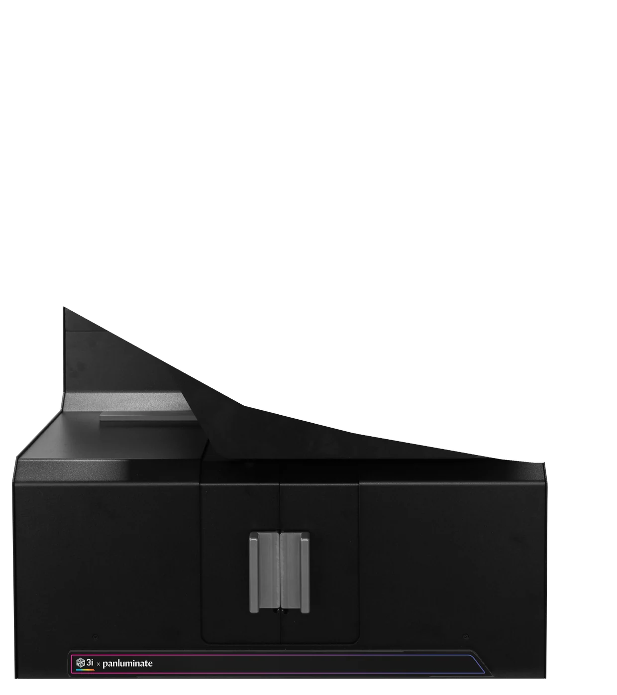
The first commercial light-sheet system purpose-built for expanded tissue. Built in partnership with 3i.
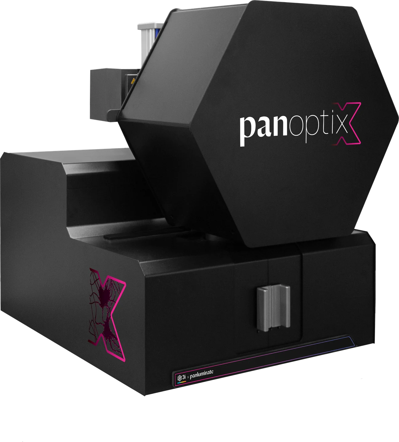
Inside panoptix
A closer look
-
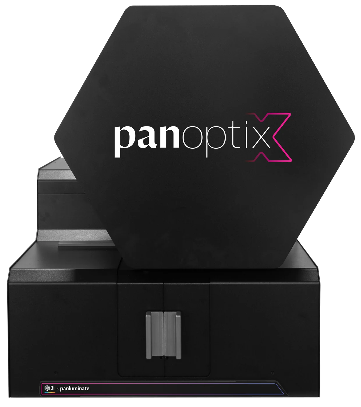
Light sheet
- Light sheet
- Axially swept lightsheet microscopy (ASLM), 420 nm sheet thickness (FWHM, 488 nm)
- Lasers
- Up to 8 lines, 405 to 785 nm, up to 1 W (wavelength dependent)
- Speed
- 0.4 Gigavoxels/s total
-
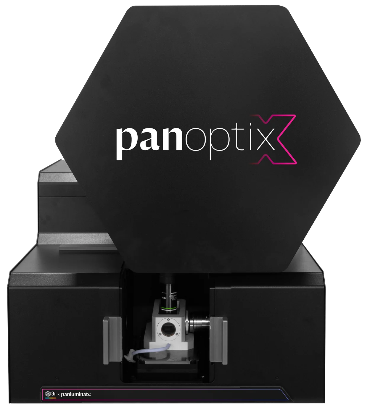
Objectives and sample chamber
- Detection
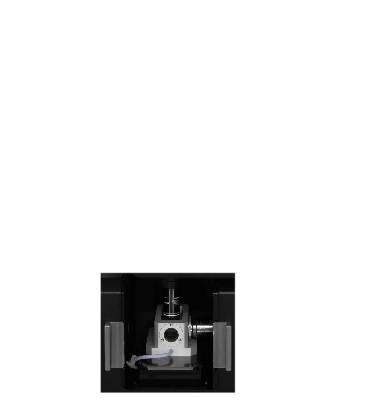
- 1.0 NA water objective, 5.45 mm WD, 48× total magnification
- Illumination
- 0.7 NA water objective, 10 mm WD
- Sample chamber
- Patented, index-matched, 10 × 22 × 5.4 mm max sample
-
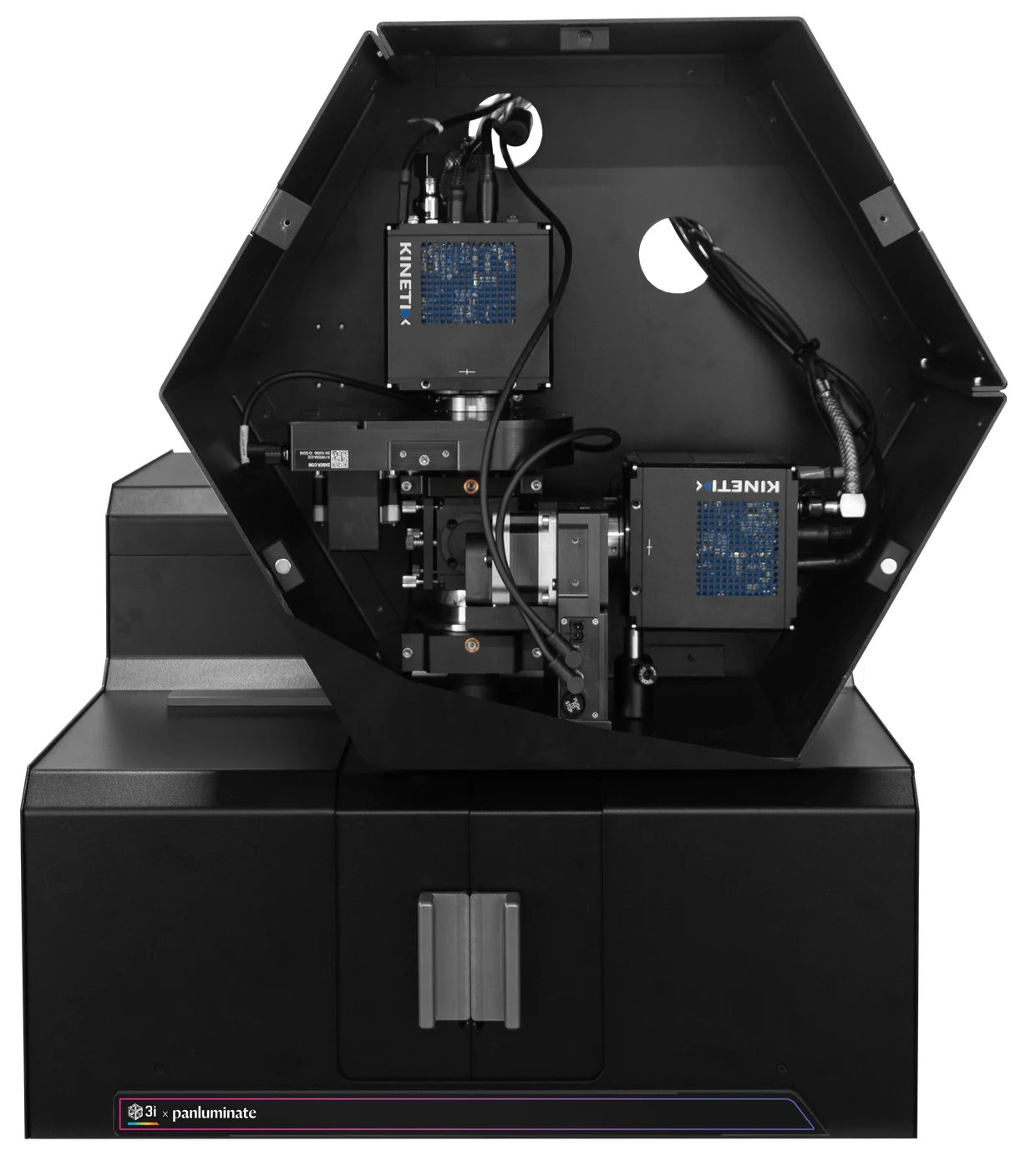
Cameras
- Cameras
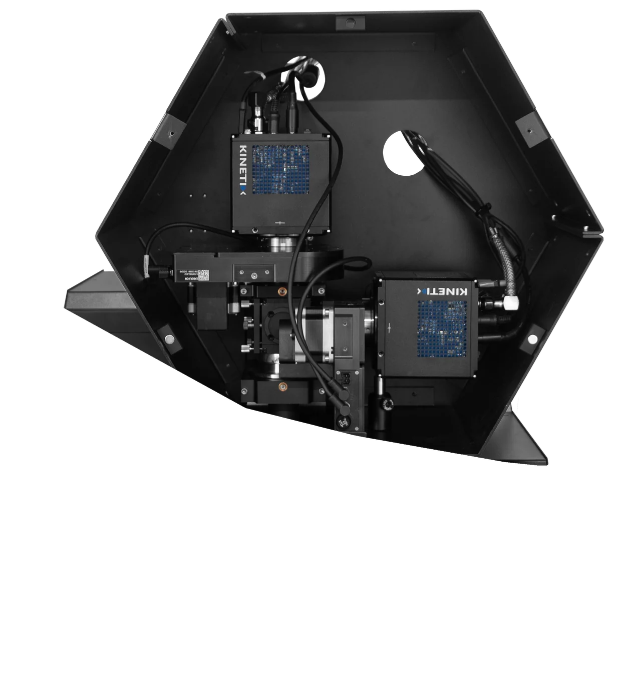
- Dual Kinetix sCMOS, simultaneous multi-color, 3200 × 3200, 135 nm pixels
Why panoptix
Expanded tissue is hard to image
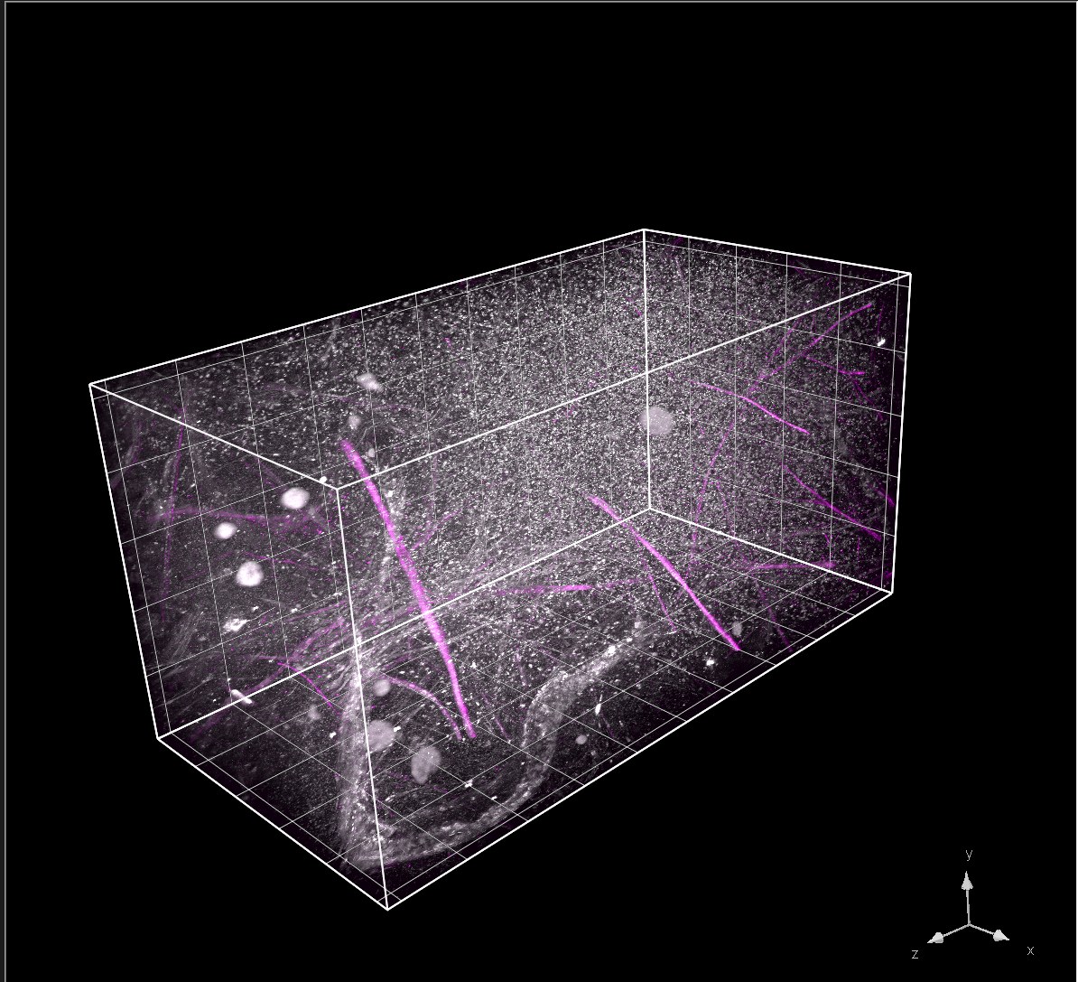
The first light-sheet system built for high-resolution expanded tissue imaging, with 2× better z-resolution than point-scanning confocal.
Specifications
panoptix at a glance
- Light sheet
- Axially swept lightsheet microscopy (ASLM)
420 nm sheet thickness (FWHM, 488 nm) - Detection
- 1.0 NA water objective, 5.45 mm WD, 48× total magnification
- Cameras
- Dual Kinetix sCMOS, simultaneous multi-color, 3200 × 3200, 135 nm pixels
- Illumination
- 0.7 NA water objective, 10 mm WD
- Lasers
- Up to 8 lines, 405 to 785 nm, up to 1 W (wavelength dependent)
- Sample chamber
- Patented, index-matched, 10 × 22 × 5.4 mm max sample
- Speed
- 0.4 Gigavoxels/s total